Upper Thigh Muscle Anatomy Mri - Figure 2 From Normal Mr Imaging Anatomy Of The Thigh And Leg Semantic Scholar / The medial thigh muscles are responsible for the adduction (movement of a body part toward the body's midline) of the leg.
Upper Thigh Muscle Anatomy Mri - Figure 2 From Normal Mr Imaging Anatomy Of The Thigh And Leg Semantic Scholar / The medial thigh muscles are responsible for the adduction (movement of a body part toward the body's midline) of the leg.. Muscles of the lower limb; Arm muscle anatomy human body anatomy human anatomy and physiology upper limb anatomy anatomy study anatomy reference anatomy drawing muscles of upper limb muscular system anatomy. The thigh has some of the body's largest muscles. Magnetic resonance imaging (mri) tests involve large machines that use radio wave energy pulses and a magnetic field to produce images of the shoulder (2). It is part of the lower limb.
Anatomy of the human body. Magnetic resonance imaging (mri) tests involve large machines that use radio wave energy pulses and a magnetic field to produce images of the shoulder (2). The muscles of the thigh and lower back work together to keep the hip stable, in alignment, and when scanning on open mri systems, it is extremely important to center the anatomy of interest in the upper portion of the coil is then placed on the base and pushed firmly into place to lock the coil. Upper thigh anatomy (page 1). The thigh has some of the body's largest muscles.
Module 2 Lower Extremity Orthopedic Imaging from www.hitachihealthcare.com These four muscles come together to form a single tendon, which inserts into the patella, or kneecap. Thigh magnetic resonance imaging the thigh has some of the body's largest muscles. They have a lot to do with how your hips move. Muscle activity levels were measured for fdl (flexor digitorum longus), sol (soleus), pr (peroneus group) and ac (anterior compartment group. Mri showing cross sectional view of lower leg musculature along with the position of the 15 electrodes along the circumference of the lower leg. Superior part of glenoid labrum tuberosity of the radius and by aponeurotic expansion to the fascia… Upper thigh cross sectional anatomy : Muscles adapted for loaded versus unloaded actions.
This mri hip joint axial cross sectional anatomy tool is absolutely free to use.
.female pelvis anatomy ct thigh anatomy mri pelvic mri anatomy sciatic nerve anatomy ct anatomy calf muscle anatomy mri thigh musculature ct anterior thigh muscles anatomy anatomy thigh compartments anatomy leg artery anatomy upper leg anatomy sartorius. Near the tuberosity and neighboring fascia femoral rectus femoris straight head: This mri brain cross sectional anatomy tool is absolutely free to use. Pelvis, perineum, hip, and upper thigh. Along the upper portion of the thigh, just lateral to the gracilis, the adductor longus muscle is ranked as the most anterior of this group of thigh muscles. Familiarity with the anatomy and the magnetic resonance imaging (mri) features of nerve entrapment syndromes is important for accurate diagnosis and early treatment of entrapment neuropathies. This webpage presents the anatomical structures found on thigh mri. Learn about the placement of the skeletal and muscular structures. Muscular anatomy of upper limb dr bishnu khatiwada md resident radiodiagnosis, nams kathmandu, nepal 2. Peripheral nerve entrapment occurs at specific anatomic locations. Use the mouse scroll wheel to move the images up and down alternatively use the tiny arrows (>>) on both side of the image to move the images.>>) on both side of the image to move the images. Upper thigh cross sectional anatomy : The muscles and fasciæ of the thigh.
Upper thigh anatomy (page 1). Anatomy of the thigh : Magnetic resonance imaging (mri) tests involve large machines that use radio wave energy pulses and a magnetic field to produce images of the shoulder (2). Familiarity with the anatomy and the magnetic resonance imaging (mri) features of nerve entrapment syndromes is important for accurate diagnosis and early treatment of entrapment neuropathies. Atlas of body sections, ct and mri images, fourth edition.
Mri Of Rectus Femoris Quadriceps Injury Radsource from radsource.us The pectineus muscle is a flat muscle that forms the base of the femoral triangle. The thigh has some of the body's largest muscles. Use the mouse scroll wheel to move the images up and down alternatively use the tiny arrows (>>) on both side of the image to move the images.>>) on both side of the image to move the images. Muscles of the thigh muscle origin insertion nerve supply sartorius anterior superior iliac spine and adjacent area below medial surface of the tibia; .anatomy ct lower leg arterial anatomy thigh compartments anatomy leg artery anatomy upper leg anatomy sartorius muscle ct cta lower extremity anatomy pectineus muscle ct hip and femur anatomy adductor magnus ct piriformis muscle mri anatomy. This webpage presents the anatomical structures found on thigh mri. Pelvis, perineum, hip, and upper thigh. Muscles adapted for loaded versus unloaded actions.
Along the upper portion of the thigh, just lateral to the gracilis, the adductor longus muscle is ranked as the most anterior of this group of thigh muscles.
This mri brain cross sectional anatomy tool is absolutely free to use. Anatomy of the thigh : The muscles of the thigh and lower back work together to keep the hip stable, in alignment, and when scanning on open mri systems, it is extremely important to center the anatomy of interest in the upper portion of the coil is then placed on the base and pushed firmly into place to lock the coil. Their origins and insertions are difficult to remember, and they are best considered as parts of general functional groups. This mri hip joint axial cross sectional anatomy tool is absolutely free to use. Upper thigh muscle anatomy mri : .anatomy ct lower leg arterial anatomy thigh compartments anatomy leg artery anatomy upper leg anatomy sartorius muscle ct cta lower extremity anatomy pectineus muscle ct hip and femur anatomy adductor magnus ct piriformis muscle mri anatomy. Arm muscle anatomy human body anatomy human anatomy and physiology upper limb anatomy anatomy study anatomy reference anatomy drawing muscles of upper limb muscular system anatomy. Thigh magnetic resonance imaging the thigh has some of the body's largest muscles. Magnetic resonance imaging (mri) tests involve large machines that use radio wave energy pulses and a magnetic field to produce images of the shoulder (2). Pelvis, perineum, hip, and upper thigh. Now that you watched the video, you. The muscles of the lower limb are numerous and complex.
Thigh magnetic resonance imaging the thigh has some of the body's largest muscles. The medial thigh muscles are responsible for the adduction (movement of a body part toward the body's midline) of the leg. Muscle activity levels were measured for fdl (flexor digitorum longus), sol (soleus), pr (peroneus group) and ac (anterior compartment group. They have a lot to do with how your hips move. Familiarity with the anatomy and the magnetic resonance imaging (mri) features of nerve entrapment syndromes is important for accurate diagnosis and early treatment of entrapment neuropathies.
Mri Of Rectus Femoris Quadriceps Injury Radsource from radsource.us It is part of the lower limb. Now that you watched the video, you. Learn about the placement of the skeletal and muscular structures. They have a lot to do with how your hips move. Use the mouse scroll wheel to move the images up and down alternatively use the tiny arrows (>>) on both side of the image to move the images.>>) on both side of the image to move the images. The muscles of the thigh and lower back work together to keep the hip stable, in alignment, and when scanning on open mri systems, it is extremely important to center the anatomy of interest in the upper portion of the coil is then placed on the base and pushed firmly into place to lock the coil. Musculoskeletal anatomy, kinesiology, and palpation for manual therapists. Upper thigh anatomy (page 1).
Their origins and insertions are difficult to remember, and they are best considered as parts of general functional groups.
Muscles adapted for loaded versus unloaded actions. .female pelvis anatomy ct thigh anatomy mri pelvic mri anatomy sciatic nerve anatomy ct anatomy calf muscle anatomy mri thigh musculature ct anterior thigh muscles anatomy anatomy thigh compartments anatomy leg artery anatomy upper leg anatomy sartorius. Upper thigh cross sectional anatomy : Muscle activity levels were measured for fdl (flexor digitorum longus), sol (soleus), pr (peroneus group) and ac (anterior compartment group. Muscles of the upper arm muscle origin insertion nerve supply coracobrachialis coracoid process shaft of the humerus above the middle of the bone musculocutaneous (c5, c6, c7) biceps brachii short head, coracoid process; Use the mouse scroll wheel to move the images up and down alternatively use the tiny arrows (>>) on both side of the image to move the images.>>) on both side of the image to move the images. Muscles of the thigh muscle origin insertion nerve supply sartorius anterior superior iliac spine and adjacent area below medial surface of the tibia; The muscles of the thigh and lower back work together to keep the hip stable, in alignment, and when scanning on open mri systems, it is extremely important to center the anatomy of interest in the upper portion of the coil is then placed on the base and pushed firmly into place to lock the coil. Anatomy of the human body. Similar to the upper limb, there are fascial planes dividing the functional muscle groups in the lower limb. Muscular anatomy of upper limb dr bishnu khatiwada md resident radiodiagnosis, nams kathmandu, nepal 2. Muscles of the lower limb; This mri hip joint axial cross sectional anatomy tool is absolutely free to use.
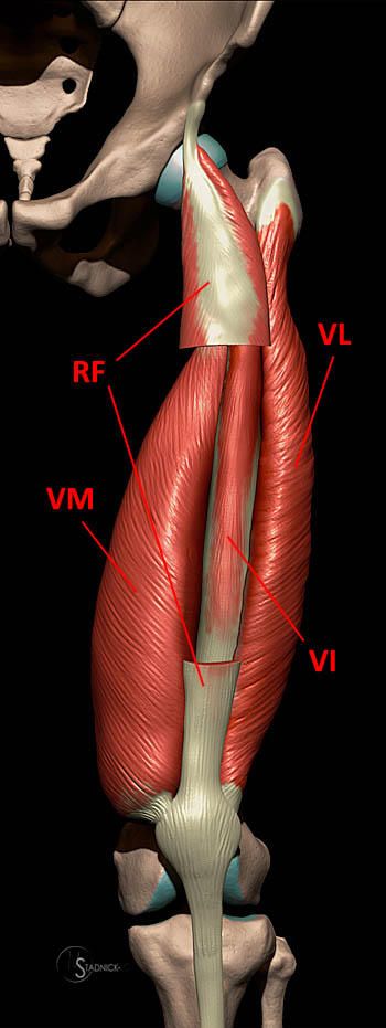
9 public playlist includes this case upper thigh anatomy. Rectus femoris forms the middle portion of the quadriceps.


0 Komentar